New study of Moyamoya - a rare disease in the brain vessels
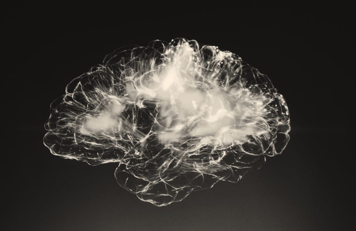
Silent damage: How Moyamoya disease shrinks the brain
Moyamoya disease is a rare but serious brain condition where important arteries deep in the brain, specifically parts of the internal carotid artery, narrow over time.
As a result, small, fragile blood vessels form on each side to bypass these blockages. While these new vessels help carry blood, they are not as effective as the original vessels.
This puts people at risk of strokes and long-term low blood flow to the brain, especially if the condition develops early in life. Over time, even without obvious strokes, this constant lack of proper blood supply may lead to permanent brain damage.
This new study aimed to find out whether such long-term reduced blood flow makes the brain shrink. The research team used high-resolution brain scans from adult patients with Moyamoya disease and compared them with scans from healthy people.
They looked closely at different brain regions and measured their volumes.
They also studied how the disease stage affected the brain and checked if some changes might be signs of long-term brain deterioration.
Who and how
The study included brain scans of 80 adult Moyamoya patients who had never undergone surgery. These were compared with scans from 129 healthy adults.
All scans were done using modern magnetic resonance imaging with very fine detail, aka high resolution.
The Moyamoya patients were classified using both imaging criteria and clinical disease scales. Among the patients, the condition affected both sides of the brain in 54 percent of cases and only one side in 46 percent.
The researchers used computer programs to measure the exact volumes of different brain areas in Moyamoya patients and compared those to healthy individuals, grouped by age.
They compared volumes of the brain's grey and white matter. Grey matter contains the cell bodies of neurons. It’s mostly found in the brain's outer layer (the cortex) and in clusters of neurons (nuclei) deep in the brain. White matter is made up of myelinated (myelin is a coat of fat that appears white) axons that act like communication cables, linking different brain regions and allowing fast signal transmission.
The volume of the cerebrospinal fluid-filled spaces, called the ventricles, was also calculated. As the brain shrinks, the areas with fluid enlarge.
They also checked whether volume loss was linked to disease severity or specific patterns of arterial blockage.
Less brain, more fluid in Moyamoya
The results were clear: people with Moyamoya had less brain volume than healthy people of the same age. The more advanced the disease, the more severe the volume loss, even in younger patients.
This was true for the overall brain and especially for the grey matter in the cortex, the part of the brain where the number of brain cells is most abundant and is responsible for high-level functions like thinking, memory, and movement.
The most affected areas included the frontal lobe, which controls planning and reasoning, and the insula, involved in emotion and awareness.
In people aged 30 to 60 with both sides affected, grey matter volume was up to 4.4 percent lower than in healthy peers.
As brain volume went down, the space taken up by cerebrospinal fluid increased—another clear sign of brain tissue loss. These fluid spaces expanded most in the same areas where brain shrinkage occurred.
Interestingly, the deeper parts of the brain, like the basal ganglia, also showed volume loss. These areas help regulate movement and cognition and are often intimately connected to the affected vessels in Moyamoya.
Even brain areas not directly supplied by the narrowed arteries, such as the cerebellum (hindbrain) and brainstem, showed reduced volume in older patients.
This suggests that not only does the disease directly affect certain regions, but it may also cause secondary damage in distant brain areas due to long-term changes in brain connectivity.
Most patients showed signs of chronic damage in the brain’s white matter, as seen on their scans, even if they hadn’t had a stroke.
Why this matters
What makes these findings especially important is that many of the Moyamoya patients had not suffered visible strokes. Their brains showed signs of wear and tear despite the absence of obvious damage or lesions on standard scans.
This supports the idea that long-standing low blood flow can cause the brain to slowly deteriorate, even in the absence of acute events.
Some parts of the brain may be more vulnerable to this slow damage. The grey matter has a higher blood supply than white matter, making it more sensitive to reduced flow.
But white matter, with fewer blood vessels, may be more prone to silent injuries, especially in the “watershed” zones where blood supply is weakest. Watershed areas are parts of the brain that lie between major blood supply zones. Because they’re at the edges of these zones, they get less blood if pressure drops, making them more vulnerable to ischemia.
Damage to white matter can interfere with communication between brain regions, as the connections between brain areas run in the white matter. That may explain some of the thinking and memory issues found in these patients.
Previous studies have suggested that patients with Moyamoya might suffer from subtle cognitive decline, even before doctors can detect clear symptoms.
The current research supports this by showing visible changes in brain structure, especially in areas tied to memory, language, and coordination.
Global brain effects from Moyamoya
Another striking finding was that patients also had brain changes far from the areas most directly affected by Moyamoya.
Brain regions like the thalamus, caudate, and hippocampus—deep inside the brain and critical for memory and motor control—showed reduced volume. These areas may suffer because they are connected to regions with poor blood supply.
When those connected areas become damaged, they may send fewer signals, leading to “downstream” degeneration. This pattern is similar to what happens in stroke patients, where not only the area of the stroke but also connected regions deteriorate and shrink over time.
The study also found changes in parts of the brain that are not usually considered vulnerable in Moyamoya, such as the cerebellum and brainstem.These regions are supplied by different arteries, which are typically not affected by Moyamoya. The damage here may be due to changes in the broader brain network rather than local blood flow issues.
This strengthens the idea that Moyamoya is not just a localized problem but can affect the entire brain over time.
Shrinkage is a sign of the silent brain damage in Moyamoya
This study helps us understand why some Moyamoya patients develop thinking or memory problems even when they haven't had a major stroke.
It shows that even in patients without visible infarcts, their brain structure is measurably different. These changes are especially clear in the grey matter, home of the brain cells, which is critical for complex brain functions.
While some regions showed thickening or thinning depending on the disease stage, the consistent finding was that grey matter and white matter shrinkage correlated with reduced blood flow and increased disease burden.
This gradual atrophy could explain the slow mental decline reported in some patients and might be used in the future to monitor disease progression and the effects of treatment.
About the Scientific Paper:
First author: Patrick Haas, Germany
Published: Brain Communications, March 2025
Link to paper: https://academic.oup.com/braincomms/article/7/2/fcaf100/8051153?login=false


Comments ()